- Diseases
- Acoustic Neuroma (16)
- Adrenal Gland Tumor (24)
- Anal Cancer (70)
- Anemia (2)
- Appendix Cancer (18)
- Bile Duct Cancer (26)
- Bladder Cancer (74)
- Brain Metastases (28)
- Brain Tumor (234)
- Breast Cancer (726)
- Breast Implant-Associated Anaplastic Large Cell Lymphoma (2)
- Cancer of Unknown Primary (4)
- Carcinoid Tumor (8)
- Cervical Cancer (164)
- Colon Cancer (168)
- Colorectal Cancer (118)
- Endocrine Tumor (4)
- Esophageal Cancer (44)
- Eye Cancer (36)
- Fallopian Tube Cancer (8)
- Germ Cell Tumor (4)
- Gestational Trophoblastic Disease (2)
- Head and Neck Cancer (14)
- Kidney Cancer (130)
- Leukemia (342)
- Liver Cancer (50)
- Lung Cancer (286)
- Lymphoma (278)
- Mesothelioma (14)
- Metastasis (30)
- Multiple Myeloma (100)
- Myelodysplastic Syndrome (60)
- Myeloproliferative Neoplasm (6)
- Neuroendocrine Tumors (16)
- Oral Cancer (102)
- Ovarian Cancer (178)
- Pancreatic Cancer (160)
- Parathyroid Disease (2)
- Penile Cancer (14)
- Pituitary Tumor (6)
- Prostate Cancer (150)
- Rectal Cancer (58)
- Renal Medullary Carcinoma (6)
- Salivary Gland Cancer (14)
- Sarcoma (238)
- Skin Cancer (300)
- Skull Base Tumors (56)
- Spinal Tumor (12)
- Stomach Cancer (66)
- Testicular Cancer (28)
- Throat Cancer (92)
- Thymoma (6)
- Thyroid Cancer (100)
- Tonsil Cancer (30)
- Uterine Cancer (86)
- Vaginal Cancer (18)
- Vulvar Cancer (22)
- Cancer Topic
- Adolescent and Young Adult Cancer Issues (22)
- Advance Care Planning (12)
- Biostatistics (2)
- Blood Donation (18)
- Bone Health (8)
- COVID-19 (360)
- Cancer Recurrence (120)
- Childhood Cancer Issues (120)
- Clinical Trials (628)
- Complementary Integrative Medicine (22)
- Cytogenetics (2)
- DNA Methylation (4)
- Diagnosis (238)
- Epigenetics (6)
- Fertility (62)
- Follow-up Guidelines (2)
- Health Disparities (14)
- Hereditary Cancer Syndromes (128)
- Immunology (18)
- Li-Fraumeni Syndrome (8)
- Mental Health (122)
- Molecular Diagnostics (8)
- Pain Management (62)
- Palliative Care (8)
- Pathology (10)
- Physical Therapy (18)
- Pregnancy (18)
- Prevention (936)
- Research (390)
- Second Opinion (78)
- Sexuality (16)
- Side Effects (616)
- Sleep Disorders (10)
- Stem Cell Transplantation Cellular Therapy (216)
- Support (408)
- Survivorship (328)
- Symptoms (182)
- Treatment (1788)
How beautiful images can advance immunotherapy
BY Julie Nagy
4 minute read | Published November 07, 2024
Medically Reviewed | Last reviewed by Sonali Jindal, M.D. on November 07, 2024
They say a picture is worth 1,000 words.
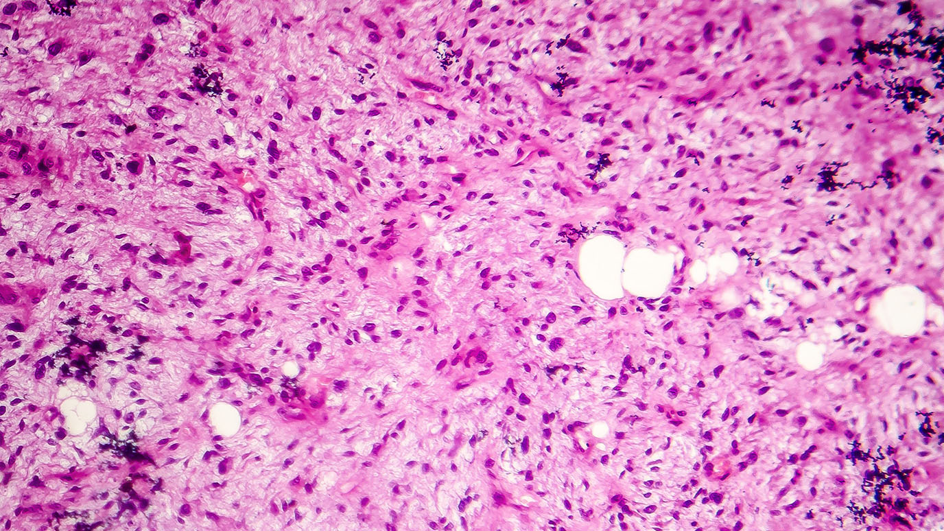
In the case of MD Anderson’s immunotherapy platform, part of the James P. Allison Institute, a picture generated by spatial omics technology provides a wealth of information to bring immunotherapy to more patients.
Immune checkpoint inhibitors can treat a variety of cancers, but patients respond differently to these combination therapies based on their unique tumor microenvironment, which is made up of many cell types, including:
- tumor cells
- immune cells
- fibroblasts
- blood vessels
- other cellular components
The immunotherapy platform uses breakthrough imaging and bioinformatics tools to paint a clear picture of the tumor microenvironment before and after immunotherapy treatment. This allows clinicians and researchers to see which cell types are present within the tumor microenvironment and how the cell types change after treatment is given. These data help researchers develop more personalized strategies to target specific cell subsets and improve responses for future patients.
We spoke with Sonali Jindal, M.D., associate director of the immunotherapy platform, to understand how these beautiful images are advancing immunotherapy treatments.
What is spatial omics, and why does it matter?
Spatial omics refers to advanced molecular techniques that analyze biological molecules within their exact location in tissue samples, creating snapshots of the tumor microenvironment. This allows researchers to see the distribution of genes and proteins being expressed, different cell-states and cell-to-cell interactions at the time the sample was collected.
The tumor microenvironment is an ecosystem that includes tumor and immune cells, blood vessels, fibroblasts and signaling proteins in and around a tumor, many of which have their own unique functions and interactions with each other, creating unique cell neighborhoods. Researchers have learned that these neighborhoods can affect whether immune cells are able to recognize and attack cancer cells and influence a tumor’s resistance to treatment.
How does the immunotherapy platform utilize spatial omics?
The immunotherapy platform aims to evaluate immune responses in patients in order to understand which specific therapies or combination therapies will need to be given so that all patients can benefit from immunotherapy. Our platform collects samples, including tumor and blood samples, for immune monitoring from patients enrolled in immunotherapy studies at MD Anderson. More than 5,000 patients have enrolled from over 100 clinical studies across various cancer types.
The samples are carefully tracked and analyzed by a collaborative network of clinicians, physician-scientists and bioinformaticians who can provide real-time monitoring of patients enrolled in clinical trials. Spatial omics is a critical part of that analysis.
How do you create these beautiful images?
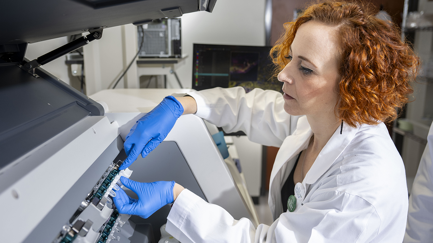
We use imaging to gain detailed information about the tumor microenvironment from these samples. Each unique type of cell has certain targets that can be tagged with a marker, usually an antibody or probe. These targets are stained using fluorescent dyes.
It works similar to “paint by number” instructions: each color corresponds to a specific target being studied. Advanced imaging tools then capture these colored sections, mapping out where each cell type or molecule is located.
For the last 50 to 60 years, it was only possible to color one or two markers on a given sample, which provided very basic information on a small scale. Even when researchers added up to nine color markers at a time, it still limited the amount of information that could be generated, given the complex environment.
Fortunately, we have new technology that significantly improves our depth of analysis. For example, CODEX (CO-Detection by indEXing) technology allows simultaneous image staining of dozens of proteins, cells and other targets in a single tumor sample. This technology also attaches a unique barcode to each antibody in order to track and quantify each target.
We want to make sure that we’re able to accurately detect the biologically relevant markers within the tumor microenvironment so that we can integrate all of the data to understand the cell-to-cell interactions and the interactions of specific markers. The data from these studies provide information about why some patients respond – or don’t respond – to treatment and which specific markers need to be targeted with new treatments so that immunotherapy can lead to clinical benefit for more patients.
How does the platform use these images to guide patient care?
The immunotherapy platform provides detailed datasets regarding the tumor microenvironment across thousands of patients, enabling researchers to focus in on specific cell subsets or molecular targets that may be important for the development of new therapies or the selection of specific patient groups for immunotherapy treatments.
We at the immunotherapy platform are very involved in studying each sample, with analyses of hundreds to thousands of molecular markers using various “omic” assays, to determine how cells are responding to treatments, and defining the specific cellular and biologic pathways that drive immune response to eliminate tumor cells.
This allows researchers to generate road maps of the different cellular interactions and neighborhoods involved in various types of cancer response or resistance to immunotherapy.
Generating and analyzing these comprehensive images is no easy task, but it is one that I consider paramount to my role. It is a big privilege for all of us to be able to do this at MD Anderson’s truly collaborative environment, where clinicians and researchers work together so closely. These beautiful pictures are a way to show the impact of immunotherapy so patients can see and understand what can be done for them. It’s all about helping patients. That is what the platform strives for.
Learn about research careers at MD Anderson.

The immunotherapy platform provides detailed datasets regarding the tumor microenvironment across thousands of patients.
Sonali Jindal, M.D.
Physician & Researcher


