- Diseases
- Acoustic Neuroma (16)
- Adrenal Gland Tumor (24)
- Anal Cancer (70)
- Anemia (2)
- Appendix Cancer (18)
- Bile Duct Cancer (26)
- Bladder Cancer (74)
- Brain Metastases (28)
- Brain Tumor (234)
- Breast Cancer (724)
- Breast Implant-Associated Anaplastic Large Cell Lymphoma (2)
- Cancer of Unknown Primary (4)
- Carcinoid Tumor (8)
- Cervical Cancer (164)
- Colon Cancer (168)
- Colorectal Cancer (118)
- Endocrine Tumor (4)
- Esophageal Cancer (44)
- Eye Cancer (36)
- Fallopian Tube Cancer (8)
- Germ Cell Tumor (4)
- Gestational Trophoblastic Disease (2)
- Head and Neck Cancer (14)
- Kidney Cancer (130)
- Leukemia (342)
- Liver Cancer (50)
- Lung Cancer (286)
- Lymphoma (278)
- Mesothelioma (14)
- Metastasis (30)
- Multiple Myeloma (100)
- Myelodysplastic Syndrome (60)
- Myeloproliferative Neoplasm (6)
- Neuroendocrine Tumors (16)
- Oral Cancer (102)
- Ovarian Cancer (176)
- Pancreatic Cancer (160)
- Parathyroid Disease (2)
- Penile Cancer (14)
- Pituitary Tumor (6)
- Prostate Cancer (150)
- Rectal Cancer (58)
- Renal Medullary Carcinoma (6)
- Salivary Gland Cancer (14)
- Sarcoma (238)
- Skin Cancer (300)
- Skull Base Tumors (56)
- Spinal Tumor (12)
- Stomach Cancer (66)
- Testicular Cancer (28)
- Throat Cancer (92)
- Thymoma (6)
- Thyroid Cancer (100)
- Tonsil Cancer (30)
- Uterine Cancer (86)
- Vaginal Cancer (18)
- Vulvar Cancer (22)
- Cancer Topic
- Adolescent and Young Adult Cancer Issues (22)
- Advance Care Planning (12)
- Biostatistics (2)
- Blood Donation (18)
- Bone Health (8)
- COVID-19 (360)
- Cancer Recurrence (120)
- Childhood Cancer Issues (120)
- Clinical Trials (628)
- Complementary Integrative Medicine (22)
- Cytogenetics (2)
- DNA Methylation (4)
- Diagnosis (238)
- Epigenetics (6)
- Fertility (62)
- Follow-up Guidelines (2)
- Health Disparities (14)
- Hereditary Cancer Syndromes (128)
- Immunology (18)
- Li-Fraumeni Syndrome (8)
- Mental Health (120)
- Molecular Diagnostics (8)
- Pain Management (62)
- Palliative Care (8)
- Pathology (10)
- Physical Therapy (18)
- Pregnancy (18)
- Prevention (936)
- Research (390)
- Second Opinion (78)
- Sexuality (16)
- Side Effects (616)
- Sleep Disorders (10)
- Stem Cell Transplantation Cellular Therapy (216)
- Support (408)
- Survivorship (328)
- Symptoms (182)
- Treatment (1788)
Spatial omics offer a deeper look at tertiary lymphoid structures in cancer
BY Devon Carter
3 minute read | Published April 12, 2023
Medically Reviewed | Last reviewed by an MD Anderson Cancer Center medical professional on April 12, 2023
Tertiary lymphoid structures are highly organized clusters of immune cells that form in non-lymphoid tissues. They’re often found at sites of inflammation, including in a variety of solid tumors. They’re made up of stromal cells as well as a diverse collection of immune cell types in a variety of cell states. Together, tertiary lymphoid structures drive an antitumor immune response. However, they’re not fully understood.
"There is still more to learn about the phenotypic heterogeneity of cells within tertiary lymphoid structures, their spatial organization patterns, intercellular communication networks and their crosstalk with cancer cells,” says computational biologist Linghua Wang, M.D., Ph.D.
Spatial technologies offer deeper insights into tertiary lymphoid structures
Traditional methods of studying tertiary lymphoid structures include histology, immunohistochemistry, multiplex immunofluorescence and flow cytometry. These methods provide the spatial location and cellular composition of tertiary lymphoid structures, but they lack the resolution and depth needed for a comprehensive understanding.
It’s like trying to understand an entire city by only knowing what certain buildings contain, without knowing where those buildings are located relative to each other.
New spatial technologies, such as single-cell spatial transcriptomics and high-plex spatial multiomics single-cell imaging, offer high-resolution spatial mapping of tertiary lymphoid structures and provide more details on cell types, functional states and cellular organization. They also provide insights into the interactions between tertiary lymphoid structures, cancer cells and other cells in the tumor microenvironment.
These new technologies not only give scientists a map of the city; they also reveal how things move and interact within it, providing a more vivid tissue context for cells.
“Without spatial profiling, we wouldn’t be able to figure out the context of cells,” Wang says. “But now we’re able to answer the questions of: ‘Where are the cells? How are they organized? Are they within the tumor bed or at the margin? Who are their neighbors?’” Wang adds.
New spatial profiling tools identify and phenotype tertiary lymphoid structures
“There’s a high demand for computational tools and frameworks that efficiently analyze spatial data at super-resolution,” Wang says. She says the demand is particularly high for tools to characterize the complex tumor microenvironment and interactions between tumor cells, immune cells and stromal cells.
Wang worked with colleagues from the University of Pennsylvania and Emory University to develop state-of-the-art computational tools that effectively integrate histology data to create an advanced spatial profile of tumor-infiltrating T cells, B cells and plasma cells both within and outside of tertiary lymphoid structures.
The tools integrate the expression data from spatial transcriptomics with the histological features of cancer. “The histology data gives us another dimension,” Wang says.
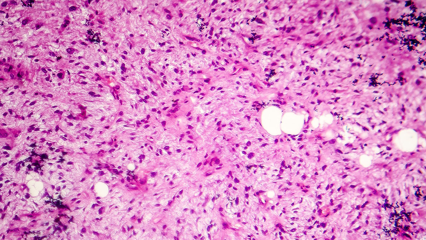
High-resolution histology images contain rich morphological information about tumors, including:
- cellular structure,
- tissue organization,
- cell size, density and shape,
- tumor grade and differentiation,
- presence of necrosis,
- invasion patterns and
- stromal characteristics.
“However, such important information is often overlooked and not fully utilized when analyzing spatial omics data,” Wang says.
At the 2023 American Association of Cancer Research (AACR) Annual Meeting, Wang will join colleagues to discuss this hot topic in cancer research. “I will be sharing the computational perspective of how B cell responses are shaped in cancer,” Wang says.
She will share the team’s experience utilizing various computational tools to detect and phenotype tertiary lymphoid structures across different cancer types.
Cell states and locations of tertiary lymphoid structures may impact immunotherapy response
In addition to several cell types being present within tertiary lymphoid structures, the cells can also vary in their cellular states. “They may contain B cells and T cells at varying differentiation and functional states, such as naïve, memory, regulatory, exhausted, and stressed cells,” Wang says. Various states of dendritic cells, fibroblasts or plasma cells that produce IgA or IgG antibodies may also be present, she adds.
The cellular composition, organization and location of tertiary lymphoid structures are essential in determining their functions. With an improved understanding of tertiary lymphoid structures within tumors, there’s hope that their full potential can be harnessed to enhance patient response to immunotherapy treatments.
“Although we are still in the early stages of utilizing spatial omics profiling, seeing in detail what’s going on with the antitumor response in tertiary lymphoid structures is a great starting place,” Wang says.

Although we are still in the early stage of utilizing spatial omics profiling, seeing in detail what’s going on with the antitumor response in tertiary lymphoid structures is a great starting place.
Linghua Wang, M.D., Ph.D.
Researcher


