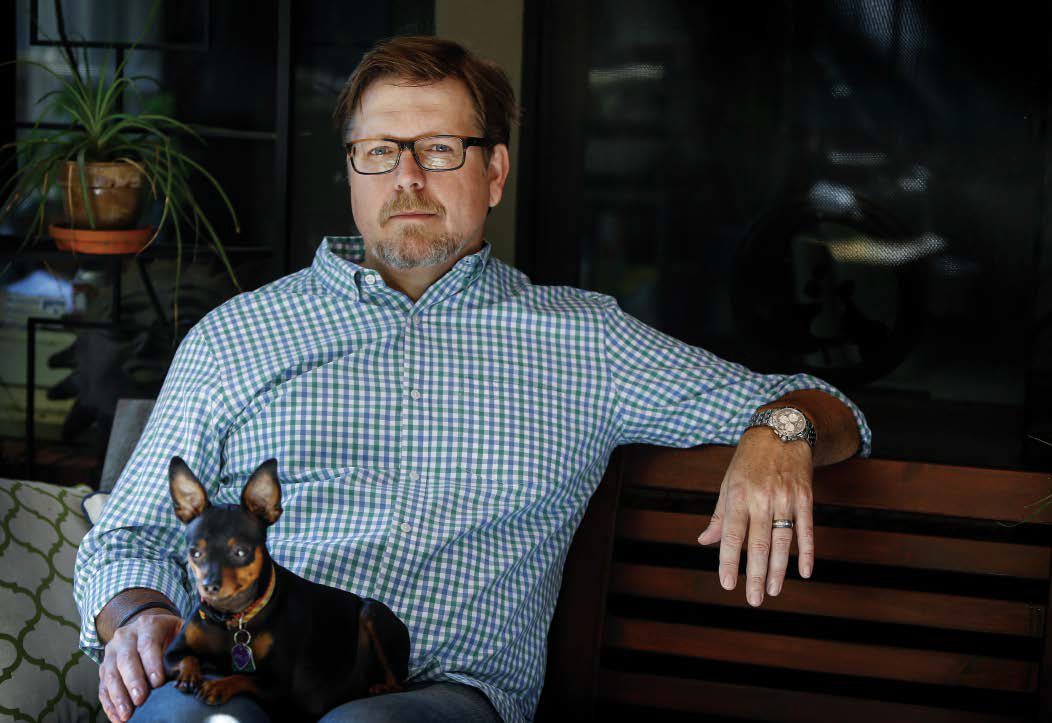
They made Jonathan Cope’s new jaw with a 3-D printer
Plastic surgery experts are using the technology to craft customized body parts that replace those damaged by cancer

Jonathan Cope lay sedated on an operating table as Matthew Hanasono, M.D., removed his cancerous lower jawbone. Meanwhile, another doctor extracted the fibula bone from Cope’s lower leg.
Using surgical tools resembling those of a sculptor, Hanasono painstakingly whittled away at the fibula, fashioning it into a new jawbone for his patient.
When he was done, he placed the new jaw where Cope’s cancer-riddled jaw had been, adjusting it till it “fit like a puzzle piece.” The arduous surgery lasted eight hours and challenged Hanasono’s skills not only as a doctor, but also an artist.

“Contouring straight leg bone into curved jaw bone is extremely challenging,” says Hanasono, a professor of Plastic Surgery. “If the fit isn’t precise, patients can become disfigured and have trouble chewing, swallowing or talking.”
He and other experts have long realized the need for a way to increase surgical precision and efficiency. Luckily, three-dimensional printing has emerged as a solution.
Eliminating the guesswork
MD Anderson is one of a growing number of cancer centers embracing 3-D printer technology to create exact replicas of body parts damaged by cancer. These replicas, or models, serve as templates to guide doctors like Hanasono as they carve and shape customized, implantable body parts out of patients’ own bones or tissues. The fibula is used to form jawbones for patients such as Cope because it’s a non-weight-bearing bone and, therefore, not essential to walking.
“Designing and making replacement body parts out of bone or cartilage once involved a lot of trial and error,” Hanasono says. “Getting accurate measurements and a good fit wasn’t easy, but 3-D printing eliminates the guesswork. Models are printed in three dimensions — length, width and height — and are precise replicas of the patient’s original jaw or other body part that needs replacing.”
Hanasono uses these models not only to create replacement body parts, but also to plan exactly how a surgery will go.
“I can take precise measurements of the model from different angles before surgery. That helps me strategize my every move,” he explains.
This surgical planning cuts down on time spent in the operating room and leads to better outcomes for patients.

A layered approach and faster recovery
Hanasono implanted Cope’s first replacement jaw five years ago after melanoma migrated from his lip to his jaw.
The surgery was performed “the old-fashioned way,” without the aid of a 3-D printed model. But radiation treatment afterward to wipe out any remaining cancer weakened Cope’s new jaw and caused it to fracture in two places.
“The docs told me my jaw was so brittle, it would continue to break,” recalls Cope, 48, a South Carolina home appliance sales manager. “I needed another one.”
Last year, Hanasono and Cope once again headed back to the operating room, but this time they had a plastic model of Cope’s jaw created with threedimensional printing.
“The technology had arrived,” Hanasono says, “and we took advantage of it.”
To create Cope’s new jaw, Hanasono and bioengineers studied a CT scan of his original jaw. Like a map, the scan guided them as they used computer-assisted design (CAD) software to produce a digital blueprint with instructions for making an exact replica.
The blueprint instructions were sent to a 3-D printer, which “printed” the jaw using plastic polymer — the “ink” used in 3-D printing. The plastic heats until it melts inside the printer, then squirts out of a nozzle resembling a miniature glue gun. After it’s expelled from the printer, the plastic solidifies and hardens into a thin layer, then more plastic is pushed out on top of it. The jaw is built one layer at a time, from the bottom up, until it’s complete.
“This process is called ‘additive’ manufacturing,” Hanasono explains, “because individual layers of material no thicker than a dollar bill are added to each other in succession, one on top of the other.”
Sometimes, prostheses are made not from plastic, but from titanium, a corrosion-resistant metal commonly used in surgery. Titanium powder is fed into the printer, then fused together with heat one layer at a time. The finished product is coated with ceramic and implanted directly in the patient.
But more commonly, the printed 3-D model, or mold, is made from plastic and is used to help the surgical team get as close to the shape and size of the original body part as possible as they carve the replacement from the patient’s bone.
Cope recovered faster from his second surgery than his first. After the first operation, he was hospitalized 12 days; after the second, just eight. But a faster recovery is only one of the advantages of 3-D printer-assisted surgery.
“My new jaw fits better, it’s stronger and more durable, and my facial symmetry has improved,” Cope says. “Overall, it’s just better. I’m working full time, golfing and enjoying life.”
And he’s looking forward to “a big steak, medium rare,” next month when doctors implant teeth in his new jawbone.
3-D Printing and the future of cancer care
The use of 3-D printing in cancer research and treatment is still in its infancy, but its implications are enormous, says Patrick Garvey, M.D., an associate professor of Plastic Surgery who uses 3-D printed models to plan his patients’ surgeries before stepping into the operating room.
“With the introduction of 3-D printing come a myriad of new applications that will someday revolutionize health care,” says Garvey, who shares a few examples:
- Surgeons are studying 3-D printed models of patients’ tumors to rehearse how they’ll remove them during surgery without harming blood vessels and nearby structures. This technology also helps medical students rehearse in a safe and forgiving simulated environment, years before they’ll operate on human patients.
- Prosthetic limbs are now being produced faster and more accurately with the technology. A CAD file that includes a person’s measurements is sent to a 3-D printer, which prints out a custom-fit limb.
- 3-D bio-printers are using living cells as “bio-ink” to print human tissue. The safety of potential new drugs can be tested on these tissues, which is less risky than testing drugs in human patients. Bio-printing also has the potential to create human organs for transplant, printed from the recipient’s own genetic matter. This will allow the new organ to precisely match the patient’s own body, and decrease the risk of rejection. Bio-printers are still experimental, but are expected to transform medicine.
- Cancer patients often have to take multiple pills each day. With 3-D printing, all a patient’s drugs could be combined in one tablet, using a printer equipped with multiple nozzles. Each nozzle would contain a different drug, and would squirt tiny amounts of each into one pill, formulated specifically for that patient.
- Patients may someday print their own drugs at home. Instead of purchasing drugs, they’ll visit an online drugstore with their digital prescription, buy the blueprint and chemical “ink” needed to make that drug, then print the drug at home on a 3-D printer that’s capable of assembling chemical compounds. The first 3-D printed pill was approved by the Food and Drug Administration last year for the treatment of epilepsy.


