Study unravels the earliest cellular genesis of lung adenocarcinoma
Findings could lead to earlier detection and intervention
MD Anderson News Release February 28, 2024
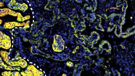
Researchers at The University of Texas MD Anderson Cancer Center built a new atlas of lung cells, uncovering new cellular pathways and precursors in the development of lung adenocarcinoma, the most common type of lung cancer. These findings, published today in Nature, open the door for development of new strategies to detect or intercept the disease in its earliest stages.
Led by Humam Kadara, Ph.D., professor of Translational Molecular Pathology and Linghua Wang, M.D., Ph.D., associate professor of Genomic Medicine, the team generated an atlas of around 250,000 normal and cancerous epithelial cells that line the lungs by studying genetic changes in each of these cells individually using a technology called single-cell sequencing.
Among the key findings of this multidisciplinary effort was the discovery and validation of a transitional alveolar cell state that harbors KRAS mutations, even in normal lung cells, and ultimately transitions to lung adenocarcinoma.
Lung cells are hijacked in transition
Alveolar cells, which are epithelial cells crucial for gas exchange inside the lung, can be grouped into two cell types. Type I cells are more common and primarily function in gas exchange, while type II cells are fewer in number and provide support for this process. In the event of injury to the lung, type II cells have inherent properties that allow them to differentiate into type I cells to replace damaged cells.
According to Kadara, this transition process can be “hijacked,” leading to a different fate for some of these transitioning type II cells.
“The large number of epithelial cells we studied, coupled with new technologies, enabled us to identify two distinct fates for type II cells,” Kadara said. “They share a common intermediate state, but one path leads to type I cells and the other progresses to tumors. Interestingly, we even found these intermediate cells in normal lung tissue and in normal regions surrounding lung cancers, and they’re stuck there. If they were transient, or quickly transitioning, we wouldn’t find as many of them, but they’re there.”
The researchers also discovered that these intermediate cells in normal tissue, which were not yet cancerous or even precancerous, had KRAS driver mutations that were not found in other cell types but matched those in the tumors of the same patients.
According to the authors, bulk sequencing previously established KRAS mutations in normal tissue. But using this new approach and other computational tools, the researchers established that these mutations were coming from one specific cell type and inferred they could be precursors to adenocarcinoma. Further analysis of this transition process is needed to fully understand the mechanisms at work.
“Leveraging multimodal and dynamic spatial imaging and molecular profiling in 2D and 3D, our team is pursuing a deeper exploration of the critical transition process during lung tumorigenesis, from normal epithelial cells to precancerous lesions and ultimately to invasive cancer,” Wang said. “The team did find that signatures of these cells were enriched in lung precancers and adenocarcinomas.”
Confirmation and next steps
To confirm their findings in vivo, the team used a model with tobacco carcinogen exposure, which creates lung damage like that incurred by cigarette smoke. They hypothesized that smoking, which is causally related to lung cancer, could stimulate the alveolar transitional state by “injuring” the lung tissue.
Not only did the models display intermediate cells before any tumor or premalignant lesion developed, but these cells persisted. The researchers found the intermediate cells persisted for six to seven months after the end of carcinogen exposure. Like their findings in the human cells, these intermediate cells had KRAS mutations and expressed signatures of KRAS activation.
Indeed, when the team generated in vitro organ models, or organoids, with these types of cells, they found the intermediate cells were highly responsive to a KRAS inhibitor.
“Our study provides unequivocal evidence that tumor cells do, indeed, arise from these intermediate cells, opening the door for new research avenues,” Kadara said. “These findings are very exciting since they suggest that KRAS inhibitors could be clinically beneficial for treatment or even interception of primitive stages of lung adenocarcinoma.”
Current efforts by the Kadara lab and collaborators, like Wang, are exploring targeting these cells with combination therapies and investigating the mechanisms of their transformation to lung adenocarcinoma including inflammation.
“These early transformations could be significantly influenced by the surrounding tumor microenvironment,” Wang said. “Obtaining a comprehensive understanding of the dynamic interactions between these cells and the immune microenvironment through in-depth spatial profiling may provide novel insights vital for early detection and the development of interception strategies.”
This work was funded by Johnson & Johnson, the National Cancer Institute (NCI) (R01CA205608, R01CA272863, T32CA217789), NCI Cancer Systems Biology Consortium (U01CA264583), NCI Human Tumor Atlas Network (1U2CCA233238), and the Cancer Prevention and Research Institute of Texas (CPRIT) (RP150079, RP220101).


