Investigating the Nature of Cancer
Though they are seldom seen by patients, pathologists take the first look at a tumor, evaluate what they discover, and help answer all-important questions for physicians and patients.


No MD Anderson patient bypasses pathology. Without the knowledge that this science provides there can be no solution — no diagnosis, no course of treatment, no cure.
Simply put, pathology is the branch of medicine that deals with the nature of disease, especially the structural and functional changes it causes in the body.
Though seldom seen by the patient, pathologists take the first look at a tumor and draw on their education, expertise and the latest technology to evaluate what they discover.
They help answer the all-important questions for clinicians and other health care providers: Does the patient have cancer? What type of cancer? What stage? What treatments might work?

Unsung heroes? To say the least
“We’re often challenged because many people simply don’t know what we do,” says Janet Bruner, M.D., professor and chair of the Department of Pathology. “Outside of medicine, people aren’t knowledgeable, but even inside of health care, many don’t have a good understanding of our role.”
Bruner attributes this to the fact that in medical school there isn’t a pathology rotation that introduces future doctors to the field. “So although head and neck surgeons also learn to deliver babies, they may never observe pathologists at work.”
Although he had extensive experience observing pathologists in all different areas, Garrett Walsh, M.D., professor in the Department of Thoracic and Cardiovascular Surgery and head of Perioperative Enterprise, agrees that this is not the case for all surgeons.
He and his colleagues in the Division of Surgery have a great deal of respect for the diagnostic abilities of MD Anderson pathologists, who have a direct impact on most surgical procedures.
MD Anderson surgeons often request a “frozen section” consult from a pathologist while in surgery. During this process a mass is surgically removed from a patient and a portion of it is frozen, cut, stained and placed on a slide for analysis — all while the patient is on the operating table. The pathologist’s analysis tells the surgeon how much of the cancer has been removed and the possibility of its recurring.
Bruner, who has served at MD Anderson since 1984 and has been chair of the Department of Pathology since 1999, says the institution is one of the most interesting places in the world to practice her specialty.
“We see very rare things here, like synovial sarcoma, a soft tissue cancer that most often occurs in the joints and tendons; things that some hospital pathologists see once or twice in a lifetime.”
Unique cases call for unique approaches
After becoming department chair, Bruner decided to specialize pathology, much like MD Anderson’s clinics are specialized.
“Our pathologists and clinicians realized that this subspecialization would create experts in our field and truly benefit the patients,” Bruner says. “Now, we have pathologists for almost every type of cancer. We have pathologists who diagnose breast cancer all day and others who look at brain tumors all day.”
Other comprehensive cancer centers also have moved to this model, and additional large hospitals across the nation are quickly following, she says.
The department’s Immunohistochemistry Lab, which uses a novel staining process to label abnormal cells, takes “specialized” to the next level.
“The histology technicians who work in this lab process about 450 slides a day,” says Kaye Barr, laboratory manager. “These are specialized readings that offer a deeper level of detail. While many of our pathologists can diagnose cancer in the liver, immunohistochemistry can tell us where the cancer originated and what type of treatment it might respond best to.”
Pam Puig, department administrator, adds that immunohistochemistry speaks to the “personalized treatment” element of the MD Anderson mission and belief that one person’s cancer is truly unique — and, therefore, so should be the course of treatment.
A testament to expertise
MD Anderson pathologists also provide consultations, the global demand for which signals that the institution is a leader in the field.
According to Sherrie Jackson, laboratory manager for Surgical Pathology Services, more than 30,000 requests for diagnoses are received each year from patients, physicians, hospitals and cancer centers around the world.
Two kinds of requests arrive — from patients who want to come to MD Anderson and, therefore, must have a pathologist review their cancer diagnosis first; or from patients or physicians who are requesting a second opinion from the pathology team, but have no intention of transferring their care here.
“Our team receives approximately 140 to 160 outside cases each day,” Jackson says. “I see it as a testament of our professionals being some of the best in the world.”
How does the future look?
As the profession progresses, the future of pathology looks bright.
“I’ve seen tremendous growth since I arrived here 26 years ago,” Bruner says. “At that time, there were 15 pathologists. By 1999, there were 30. Today we have 60. We’ve grown because MD Anderson has grown, but also because there’s a greater research component now, which is extremely important.”

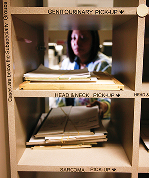
Once materials submitted through the second opinion
consultation service have been verified and processed,
Lanetha Kellum, supervisor in Surgical Pathology Services,
places them in the subspecialty service box for the
appropriate pathologist to review.
The work of Bogdan Czerniak, M.D., Ph.D., professor in the Department of Pathology and chief of the Section of Genitourinary Pathology, is one example of original research being carried out. His studies involve fundamental genetic mechanisms of bladder cancer.
The Department of Pathology also oversees analysis of fluid and tissue samples that are part of MD Anderson’s clinical trials. And when it comes to national meetings and conferences, the Division of Pathology and Laboratory Medicine consistently submits the highest number of presentations and abstracts among its colleagues. Working to continue this trend is one of Bruner’s goals.
Though MD Anderson’s Department of Pathology is one of the best, she recognizes that there is always room for improvement.
“As a department head, I want to ensure that we’re not lacking when it comes to leveraging technology to our advantage and that of our patients,” she says. “We’re no longer tied to the microscope. As a profession, we use the Internet and other digital technology to provide diagnosis and recommend treatments that are in accordance with personalized cancer care. This is where our profession is rapidly moving, and MD Anderson pathologists must aggressively move in this direction also.”
Pathology Dissected
Pathology is one of four departments in the Division of Pathology and Laboratory Medicine. Within the department there are four major areas.
Cytopathology
Cytopathology provides diagnostic laboratory service for the early detection of cancer and tumor staging. The team of cytotechnologists and cytopathologists, who are specialized to identify cancerous cells and microorganisms, play an extremely important role in cancer prevention. The Cytology Lab processes several types of samples, including gynecologic (Pap smears) and non-gynecologic samples (body cavity fluids, urines, bronchial and peritoneal washings) and fine needle aspirations (FNAs) from various body sites.
The Pap smear is the most well-known screening test for cervical cancer, according to Shobha Patel, laboratory manager for cytopathology. This technique was developed in 1940, and the test helped reduce the death rate from cervical cancer by 70% over the next 50 years. The recent addition of molecular genetic testing for high-risk human papillomavirus (HPV) in 2003 has helped triage the treatment of women at risk for cervical cancer.
Introduced in 1984, FNA is a cost-effective and minimally invasive procedure that has revolutionized several aspects of patient care, such as its use with endoscopic bronchial ultrasound in the staging of lung cancer patients, thereby decreasing the need for mediastinoscopy and open lung or lymph node biopsy.
Histopathology
Histopathology laboratories provide the technical support for processing tissue specimens submitted to the department from surgery or from the various outpatient clinics. These specimens must be grossly identified and described, and then put through a process of fixation, embedding in paraffin and finally, cutting and staining prior to delivery to the pathologists for review and diagnosis
The frozen section service aids surgeons during their cases. A specimen can be taken from a patient, prepared on a slide within 20 minutes or less and diagnosed by a pathologist, all while the patient is on the operating table. This reading helps the surgeon determine if enough of the cancerous mass has been removed and the chances of the disease recurring in one or more places. Material submitted for permanent sectioning and review is available for the pathologist within days of the patient’s surgery and provides a comprehensive interpretation of all specimens removed during the surgery.
Immunocytochemistry
The Immunocytochemistry Laboratory processes requests for ancillary staining of specific markers in tissue. The results of these stains are interpreted by the pathologist and used to differentiate diagnoses or to provide prognostic information to help determine treatment plans for patients.
Surgical Pathology
Surgical Pathology Services includes support services that are responsible for receiving, logging and tracking material received from outside facilities requesting pathology review, as well as processing requests for specialized molecular and genetic testing on pathology specimens.


