Breast Imaging: Mammography
Diagnostic Imaging Procedures
Before a patient arrives for an imaging exam, our diagnostic imaging providers review the orders for every CT, MRI and PET exam to ensure we’re conducting the most valuable study. Unlike most imaging centers, which use generic imaging protocols, our radiologists can access a patient’s records and prior imaging studies and then work with the primary provider to design an exam that answers specific clinical questions. This allows us to produce relevant, high-value, oncology-focused reports.
Our team of 21 board-certified imaging physicists continually updates and customizes our machines and imaging modalities. They design custom studies to improve cancer detection and routinely collaborate with leading companies to develop the latest imaging technology.
Subspecialized radiologists with expertise in oncology are available all day, every day to read our patients’ studies. Our radiologists ensure imaging reports are readily available to MD Anderson providers as well as to patients and referring providers (based on patient preference) via MyChart.
Diagnostic imaging clinics
In addition to offering imaging services at our Texas Medical Center locations, MD Anderson also operates imaging clinics in Bellaire, League City and West Houston. These locations offer convenient access, free parking and quick turn-around times. Learn more about our Diagnostic Imaging Clinics.
Contact us
New patients without a referral should visit our appointments page.
New patients who have an imaging referral from an outside health care provider to MD Anderson should call 346-639-2582.
Existing patients who need to schedule or reschedule imaging exams should call their home clinic or care center.
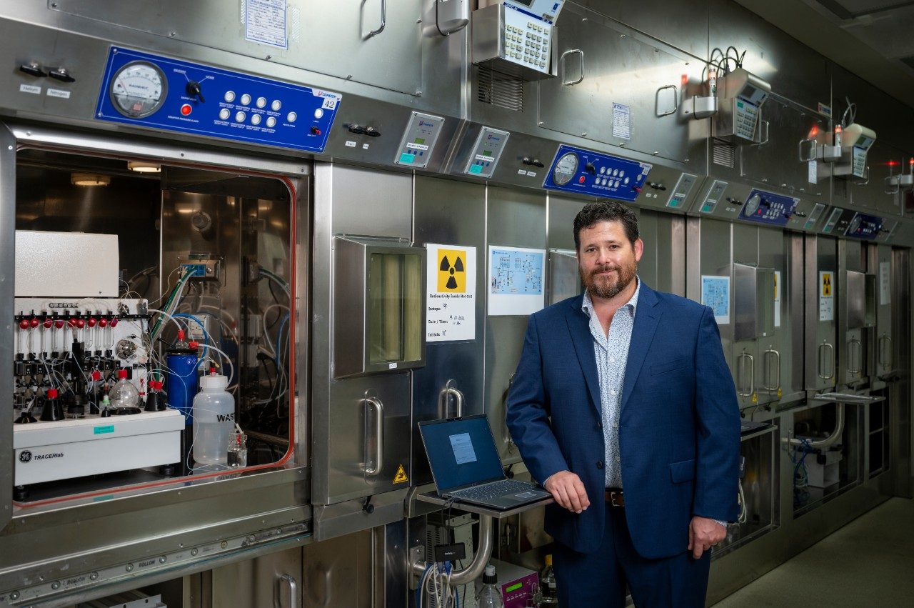
What is theranostics?
Common diagnostic imaging procedures
Breast imaging
Breast imaging captures images of breast tissue by combining multiple imaging technologies, such as mammography (the use of x-rays), ultrasound and MRI procedures.
Learn more about breast imaging on our Mammograms and Breast Examination page.

CT (Computer Axial Tomography) scan
A CAT (computerized axial tomography) scan, also known as a CT scan, uses an x-ray machine to take several pictures from different angles, providing a highly detailed image. Some CT scans require contrast to enhance the image quality. Patients may be given contrast to drink or have it administered through an IV prior to a scan. Some areas of the body that are examined with a CT scan are the chest, the nervous system and musculoskeletal systems. MD Anderson offers weekend appointments for those needing a CT.
Learn more about what to expect and how to stay safe during your CT scan.

Computerized Tomography (CT) Scan
Clinical nuclear medicine
Clinical nuclear medicine uses a small amount of radioactive tracers to indicate the presence of disease within specific organs. This imaging technique helps reveal the concentration and location of the disease. Exams performed include bone scans, bone mineral density, thyroid cancer study and more.

Clinical Nuclear Medicine: Bone Scan
Fluoroscopy/Radiography
Fluoroscopy/radiography utilizes x-rays to take a wide variety of images that form a live look at internal organs. This type of imaging is common for pediatric patients. Fluoroscopy can also be used to help guide the placement of medical devices inside the body. The spine, chest, pelvis and more are evaluated with this method.

Fluoroscopy

Lumbar Puncture (Spinal Tap) Under Fluoroscopy
General/Body ultrasound
General/body ultrasound operates with high-energy sound waves that bounce off internal tissues and organs and produce echo patterns. The echo patterns create a picture referred to as a sonogram, which can be seen on the ultrasound machine. A biopsy may also be performed during an ultrasound. Ovarian screening, pelvic bleeding and abnormal blood work are a few reasons to perform an ultrasound.

General Ultrasound
MRI (Magnetic Resonance Imaging)
MRI (magnetic resonance imaging) uses magnetic fields and radio waves, rather than radiation, to generate pictures of the body’s soft tissue and organs. Contrast may be added into the body to enhance the images. Metal outside the body, such as jewelry, must be taken off, while metal inside the body, such as surgical implants, must be removed before a scan. An MRI can be used to image the head, spine, abdomen and other body parts. MRI is available during the weekend at some MD Anderson locations.
Learn more about what to expect and how to stay safe during your MRI.

Preparing for an MRI scan at MD Anderson
PET (Positron Emission Tomography) scan
PET (positron emission tomography) scan is a technique in which a small dose of radioactive sugar is injected into a patient. A scanner shows where the sugar is being distributed, allowing for the creation of an image. The pictures can help radiologists find cancer cells in the body. Tests are conducted for PET oncology and neurology.
X-ray
X-rays are the most common way doctors attain images of the inside of the body. They use low doses of high-energy radiation that travel through the body. Radiography refers to the use of x-rays. Radiologists can spot abnormal areas in x-ray images that may indicate the presence of cancer.

X-ray: Bone Density Test
Questions About Radiation Exposure?
The benefits of diagnostic imaging are real and immediate while the risks are minimal.
Cancer Screenings
request an appointment online.
Help #EndCancer
Give Now
Donate Blood
Our patients depend on blood and platelet donations.
Shop MD Anderson
Show your support for our mission through branded merchandise.